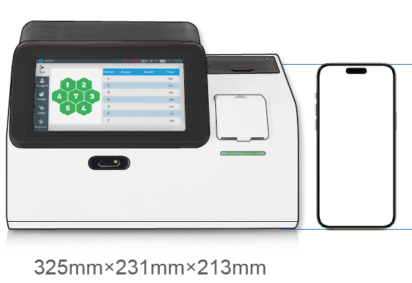
Lab-Quality Thrombosis Testing with Poclight D-Dimer POC CLIA

1.  Thrombosis: An Overlooked Threat
 

The International Society on Thrombosis and Haemostasis (ISTH) established World Thrombosis Day (WTD) on October 13 to raise awareness of the global health threat.

Every year, 1 in 4 people worldwide die from conditions related to thrombosis, the underlying cause of the top three cardiovascular killers: heart attack, stroke, and venous thromboembolism (VTE). - World Thrombosis Day website (2025)
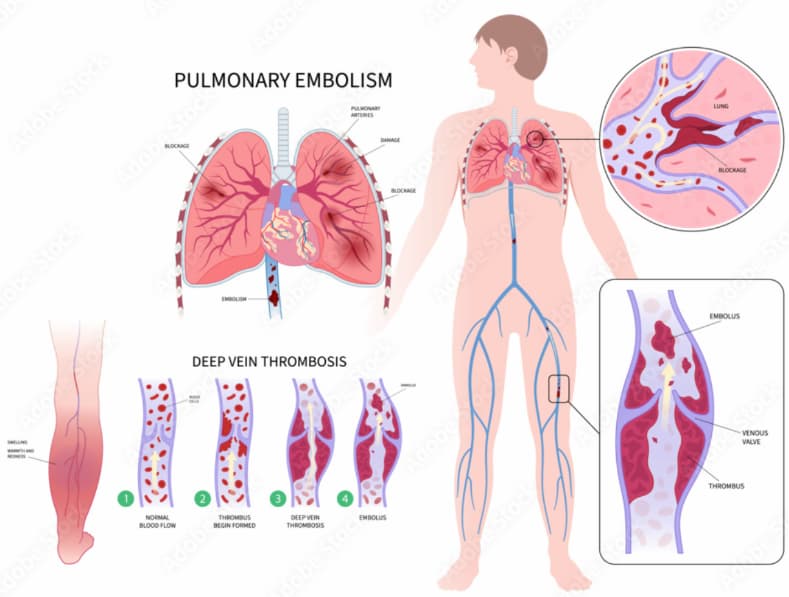
Venous Thromboembolism (VTE)


According to WHO data (2001–2023), the global age-standardized mortality rate of pulmonary embolism fell from 3.49 to 2.42 per 100,000. However, rates are rising in some lower-middle-income regions, underscoring the need to strengthen diagnostic capacity. Rapid and accessible POC testing helps bridge this gap where laboratory resources are limited.


2.  D-Dimer Testing Essentials


D-dimer is a specific fibrinolysis marker and key laboratory indicator of thrombus formation and breakdown. It rises within 2 hours of clot formation, has a 7-8 hour half-life, and remains stable in blood samples for 24-48 hours. These characteristics ensure its crucial role in diagnosing and monitoring thrombotic diseases.

Its clinical applications may include the following:

● Early Detection of Thrombosis: D-dimer testing is widely used to help rule out venous thromboembolism (VTE) in patients with suspected thrombotic events, particularly when combined with clinical assessment. Rapid detection supports prompt management decisions.

● Guiding Further Assessment: Prompts additional imaging or diagnostic tests.

● Supporting Anticoagulation Decisions: Helps guide initiation, adjustment, or duration of anticoagulant therapy.


Test Combination


Item

Clinical Use / Recommendation

D-dimer Test Kit

Helps rule out suspected DVT or PE (when combined with clinical probability assessment)

D-dimer + NT-proBNP Test Kit

Assists in differentiating PE from heart failure in patients with acute dyspnea

D-dimer + NT-proBNP and/or Troponin I

Helps rule out PE and assess PE prognosis

Note: D-Dimer levels can rise due to infection, inflammation, surgery, or pregnancy. It should always be interpreted alongside clinical findings and imaging.


3.  Poclight D-Dimer Solutions
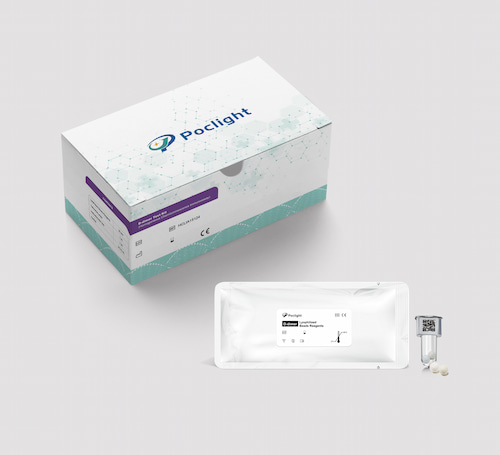
 

D-Dimer Test Kit

Highly cost-effective, Poclight Biotech’s point-of-care D-dimer Test Kit solutions deliver results within 5 minutes, supporting prompt assessment and care.


Our Stand-out features:


1)  Small Sample Volume & Rapid Results

Only 50 μL of serum or plasma needed, with 5-minute turnaround, ideal for fast clinical decisions.

Parameter

Sample type

Assay duration

Sample volume

Cut-Off value

Measuring range

D-dimer

Plasma

5min

50 μL

0.5 mg/L, Higher D-dimer levels suggest clotting activity

0.25~20 mg/L


2)  Freeze-dried reagents: no cold chain, 18 months shelf life, simplifying storage and transport.

3)  High accuracy and excellent precision (CV < 5%)
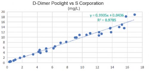
 

DDimer Performance


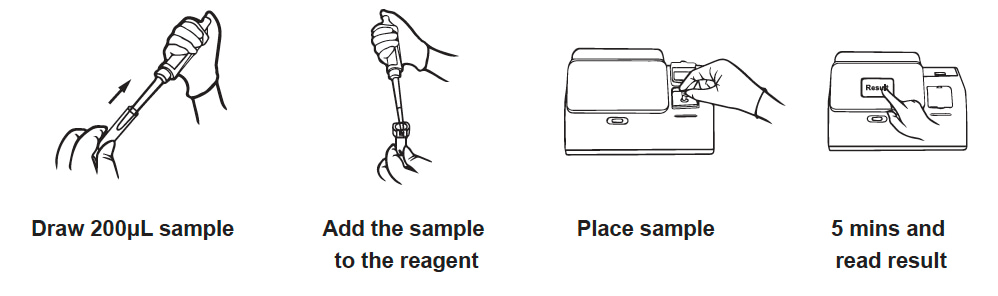
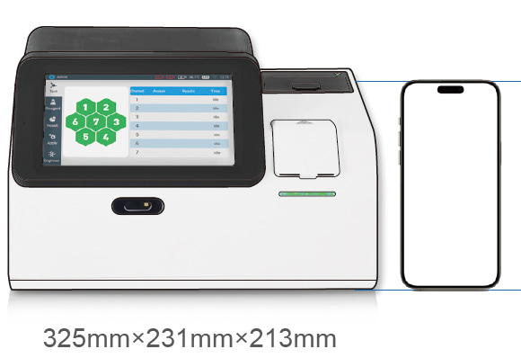
4)  Applicable with the C5000 POC CLIA Analyzer: 3-Step Operation, Maintenance-Free
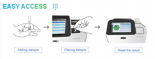
C5000 Operation


Contact us to learn about local distributors and pricing options for Poclight D-dimer solutions.
Top

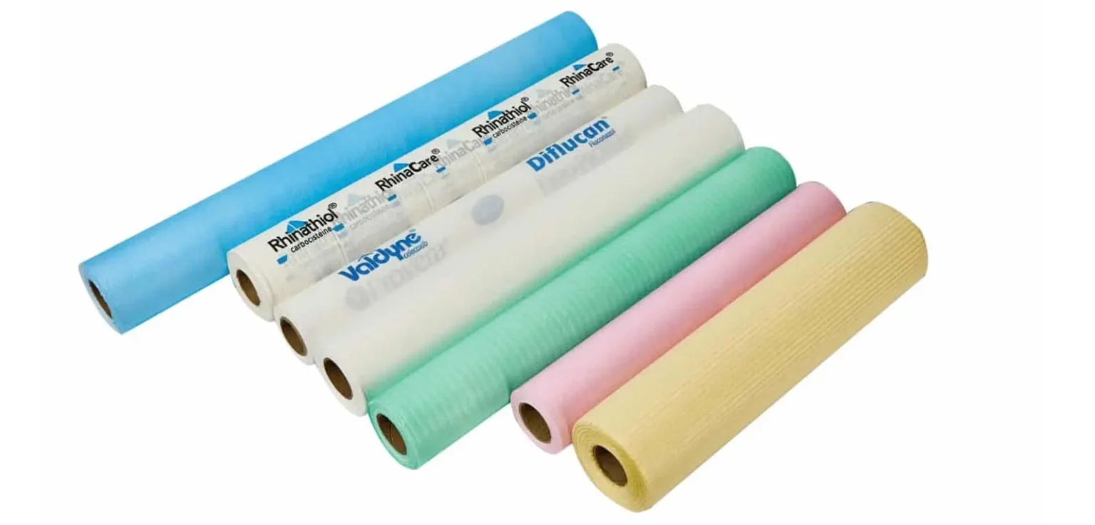
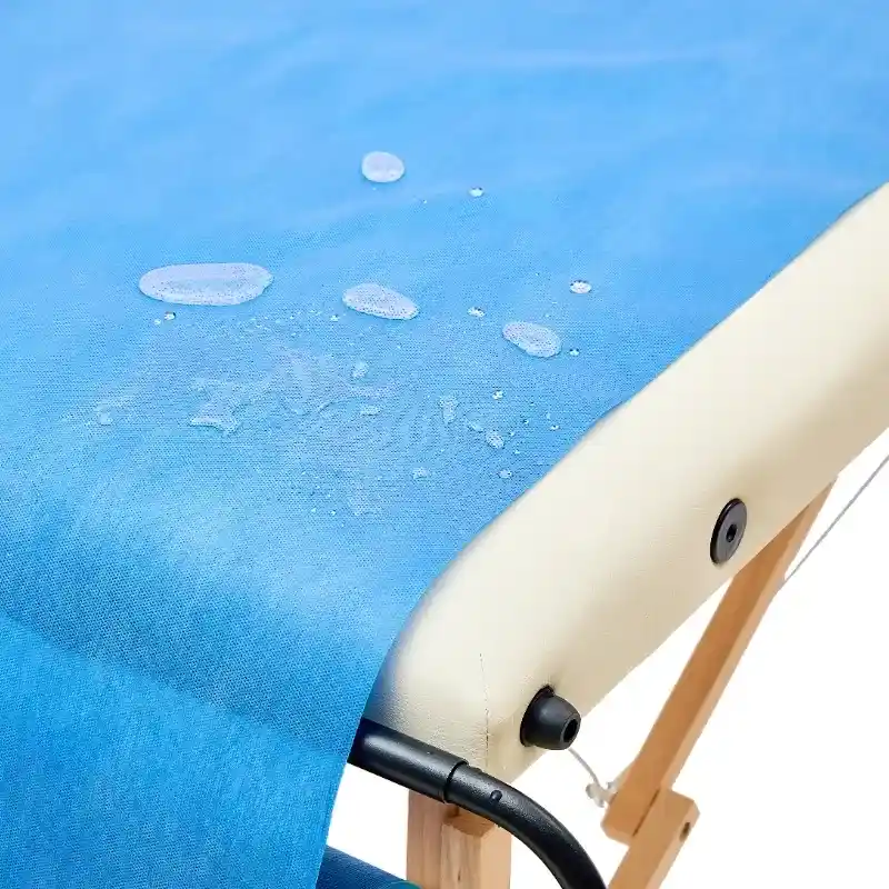
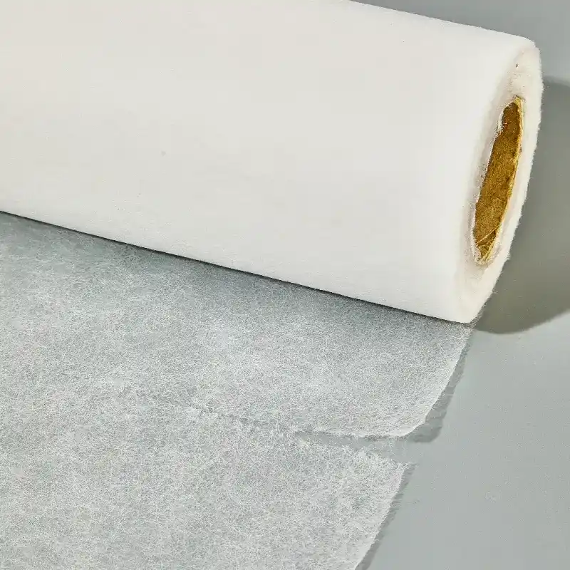
Disposable Paper Bedsheet Roll Hygienic Medical Couch Rolls for Clinics

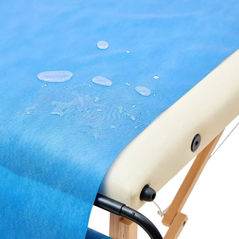
A clean, comfortable exam experience starts with the little details — and a high-quality Disposable Paper Bedsheet Roll makes a big difference. Clinics, spas, and mobile healthcare teams rely on disposable sheets to maintain hygiene between patients while keeping setup and cleanup quick. Designed for single-use convenience, these rolls help reduce cross-contamination risks and improve workflow in busy exam rooms.


For practitioners who need reliable, professional coverings, options such as Medical Couch Rolls and the Examination Couch Cover Roll are engineered to fit standard couches and trolleys, tear cleanly at perforations, and provide a soft, skin-friendly barrier. Good rolls balance strength and softness, resist minor fluids, and are available in a range of widths and ply to suit consultations, procedures, and physiotherapy sessions — all while being lightweight to store and simple to replace between patients.


At our factory we combine practical design with production discipline. With over 20 years’ experience manufacturing disposable medical disposables and supplying the global market, we offer OEM/ODM flexibility, consistent quality control, and competitive lead times. Customers benefit from tailored sizing, custom printing, and small-batch sampling so you can trial products before committing to larger runs. Our teams handle logistics and documentation for international shipping, making it straightforward to stock exam rooms anywhere in the world.


Choose Telijie when you want more than a product: you get a partner. Telijie’s strengths include stable, in-house manufacturing capacity, rigorous inspection at every stage, and dedicated account managers who respond quickly to orders, samples, and after-sales requests. We support healthcare buyers with clear packaging, compliant production practices, and flexible service options to match your inventory and budget needs — helping clinics and distributors maintain high standards without hassle.

Top

Revolutionizing Platelet Function Testing with Homogeneous CLIA Technology

1.What is Platelet?

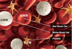
Platelets, fragments of cytoplasm derived from bone marrow megakaryocytes, play a central role in hemostasis and thrombosis. When blood vessels are injured, platelets adhere, activate, and aggregate to stop bleeding. Abnormal platelet function can lead to bleeding disorders or thrombotic events. With the widespread use of antiplatelet drugs, such as aspirin and clopidogrel, precise assessment of platelet activation function is increasingly important. Poclight’s next-generation Homogeneous CLIA (Chemiluminescence Immunoassay) technology brings a new level of accuracy and speed to platelet function testing.

.What Are Platelets


2.Platelet Function and Clinical Significance

Platelet-mediated hemostasis involves three key steps:

a. Adhesion

When the endothelium is disrupted, platelets adhere to the vessel wall via von Willebrand Factor (VWF) and collagen. This process is mediated by glycoprotein receptors CD42a/b/d (GPIb-IX-V), a critical step in platelet adhesion.

b. Activation

Platelets are activated through G-protein-coupled receptors binding to ligands such as thrombin, ADP, ATP, and prostanoids. Activated platelets change shape to a dendritic or octopus-like form.

c. Aggregation

Activated platelets are interconnected by fibrinogen and receptors CD41/61 (GPIIb/IIIa). Platelet granules release their contents into the extracellular fluid, among which CD62P (P-selectin) promotes platelet aggregation through platelet–platelet and platelet–fibrin interactions.


Platelet function

Hemostasis and thrombosis: The Key Role of Platelets 

Physiological function: The biological behavior and function of platelets are extremely complex. This includes participating in hemostasis and coagulation, maintaining vascular function, regulating inflammation, the body's immune defense, wound healing, tumor growth and metastasis, etc. Therefore, in clinical activities, the monitoring of platelet functionality is receiving increasing attention.

The average lifespan of platelets is 7 to 9 days. Senescent and dead platelets are eventually phagocytosed and processed in the spleen through the bloodstream.


Clinical Significance

  1. Thrombosis Risk Assessment:  Patients with Hypertension, diabetes,coronary, artery disease,and other cardiovascular condition, as well as those on antiplatelet or anticoagulant therapy.
  2. Pre/Postoperative Evaluation: Predicting thrombus formation and guiding prophylactic measures.
  3. Platelet Dysfunction Disorders: For example, Glanzmann’s thrombasthenia (CD41/61 deficiency) and Bernard-Soulier syndrome (CD42a/b/d deficiency).
  4.  Antiplatelet Therapy Monitoring: Supporting decisions for drugs like clopidogrel, ticagrelor, and prasugrel.
  5. Various detection functions:


Defect of platelet momoprotein receptor

CD61(GP IIIa)

CD(GP IIb)

CD42a(GP I X)

CD42b(GP I b)


Platelet Activation

CD62P(P

selectin)

CD61(GP IIIa)

CD61

Antibody(PAC-1)

CD45(White blood cell platelet aggregation)

Platelet Function

AA-CD62P

ADP-CD62P

…


3. Advantages of Poclight Platelet Activation Function Test

Feature

Description

Accurate & Fast

Quantitative results in just 5 minutes, enabling rapid clinical decisions.

Portable & Minimalist

One-step sample addition, maintenance-free, suitable for point-of-care testing.

True & Reliable

Technical details to be provided by the team to support this claim.

Quality Assurance

Supports quality control products and continuous QC checks, ensuring reliable results.

reagent testing

Technical Highlights:

Employs Homogeneous CLIA technology to detect multiple platelet surface markers (CD41, CD42, CD61, CD62P) for comprehensive assessment.

Evaluates the full spectrum of platelet function—adhesion, activation, and aggregation—delivering more comprehensive and physiologically-relevant results.


4. Compatible Devices

The Poclight Platelet Activation Function Test is fully compatible with Poclight latest C5000 Analyzer, offering seamless integration into clinical and point-of-care settings. Key compatibility features include:

  1. Plug-and-Play Operation: Simple one-step sample addition for immediate testing.
  2. Portable Analyzer Integration: Works with compact, maintenance-free devices suitable for outpatient clinics or bedside monitoring.
  3. Future Expansion: Designed to support upcoming analyzer models based on the Homogeneous CLIA platform.
  4. Note: For detailed technical specifications and compatibility with your laboratory instruments, please contact our technical support team at overseas@poclight.com.

 Chemiluminescence immunoassay solution


5. Clinical Applications

  1. Outpatient Thrombosis Risk Screening: Quickly assess high-risk patients and guide antiplatelet therapy.
  2. Pre/Postoperative Monitoring: Track platelet activity changes to optimize surgical management.
  3. Antiplatelet Drug Efficacy Evaluation: Support individualized medication decisions and reduce bleeding or thrombotic events.
  4. Diagnosis of Platelet Dysfunction Disorders: Provide molecular-level insights for rare platelet diseases.


6.Influencing factors

  1. Genetic factors: Genes determine each person's basic platelet activity and their response to drugs.
  2. Drugs: Antiplatelet drugs, NSAIDs (such as ibuprofen), certain antibiotics, antidepressants, etc., can all affect their function.
  3. Disease status: Chronic diseases such as diabetes, kidney disease, and autoimmune diseases are often accompanied by high platelet reactivity (easier to form blood clots).
  4. Lifestyle:

Diet: Omega-3 fatty acids, garlic, ginger, certain vitamins (such as VC, VE) may inhibit platelet function.

Smoking and drinking: Smoking activates platelets, while moderate drinking may inhibit them.

Physiological conditions: Stress, exercise, and aging can all increase platelet activity.


7.Future Outlook

Platelet activation function testing is evolving towards speed, portability, standardization, and intelligence. Poclight’s Homogeneous CLIA platform not only shortens testing time but also delivers reliable, high-quality data, supporting precise assessment of bleeding and thrombotic risk. With ongoing technological innovation, this approach will play an increasingly vital role in precision medicine and individualized patient care.


8.Chemiluminescence resonance energy transfer(CRET)

Detection principle:

CRET cell technique utilizes the molecules on the platelet membrane to specifically fix and bind to the corresponding antibody 1-labeled DNA1, antibody 2-labeled DNA2, and AE-labeled DNA3 to form complexes. By detecting the light intensity via CRET, the expression levels of various antigens and receptor molecules in the sample can be detected.

Project Reference Interval Project

Reference Interval

CD62P

1.04-4.52%

CD42a

82.65-100%

CD61抗体

0-10%

CD42b

81.86-100%

CD61

60-92.13%

CD45

12.95%-46.04%

CD41

90.7-99.0%


Detection method:

  1. Nucleic acid technology is integrated into immunodiagnosis, leveraging the proximity efffect of nucleic acids takes effect.
  2. The properties of graphene oxide are strategically utilized, eliminating the need for magnetic beads and their associated separation and washing steps, and simplifying the fluidic path.
  3. The introduction of enzymatic cycling amplification technology enhances detection sensitivity by 1 to2 orders of magnitude, improving overall performance while simultaneously simplifying the process.


Advantage:

CRET compensates for the drawbacks of flow cytometry and enables:

  1. Fast, reliable detection in just 5-10 minutes.
  2.  A simple and minimalist operational procedure.
  3. Low-cost testing with no hidden fees.
  4. Easy adoption in all levels of hospitals and departments.
  5. Accurate in vivo status: Not affected by platelet aggregation or in vitro activation issues. 


Contact Us

E-mail: overseas@poclight.com


Top

Understanding Optical Coherence Tomography Technology in One Article.

I. What is OCT? A Core Metaphor


Let’s understand OCT through a vivid analogy:

Imagine a fresh sandwich. A regular camera can only capture its top view (a two-dimensional photo). OCT, however, acts like a precise “optical scalpel.” It can slice the sandwich layer by layer without damage, capturing ultra-high-resolution images of each component (bread, vegetables, meat slices). These cross-sectional images are then reconstructed into a three-dimensional model.

In the eye, this “sandwich” is your retina—the light-sensitive tissue at the back of the eyeball. OCT generates cross-sectional images of the retina’s microscopic structure without any incisions or contact, achieving micrometer-level precision (1 micrometer = 0.001 millimeters)—far finer than a strand of hair!


II. How Does OCT Work?

OCT operates on a principle similar to ultrasound, but instead of sound waves, it uses near-infrared light.

  1. Emission: The device emits a safe, low-energy near-infrared light beam toward your eye.
  2. Reflection: This light passes through the eyeball and reflects back from different layers of the retina. Each tissue layer reflects light signals with distinct intensities and time delays.
  3. Analysis: The device’s core interferometer precisely measures the differences between these reflected signals and the reference light (“interference” phenomenon).
  4. Imaging: A computer analyzes and processes the countless captured light signal points, instantly constructing detailed 2D or 3D images.

    III. What Can OCT Detect? Which Eye Conditions Does It Diagnose?
    OCT is a powerful tool for examining the macula (the central part of the retina responsible for sharpest vision). With it, doctors can:
  5. Precisely measure thickness: Determine whether the retina has thickened (edema, hemorrhage) or thinned (atrophy).
  6. Identify minute lesions: Detect fluid accumulation, abnormal membranes, and microbleeds invisible to the naked eye.
  7. Monitor disease progression dynamically: Evaluate treatment efficacy and guide subsequent therapeutic decisions.

    It is primarily applied in the diagnosis and management of these major blinding eye diseases:
  8. Age-related macular degeneration (AMD)

    Wet AMD: OCT clearly visualizes abnormal subretinal vessels and fluid accumulation, serving as the gold standard for diagnosis and determining the need for intravitreal injections.

    Dry AMD: OCT monitors for macular thinning (geographic atrophy) and the deposition of drusen.
  9. Diabetic Retinopathy & Diabetic Macular Edema (DME)

    OCT is the most sensitive tool for detecting DME (intraretinal fluid accumulation), precisely guiding laser or anti-VEGF drug injection treatments while objectively evaluating their efficacy.
  10. Glaucoma

    OCT accurately measures the thickness of the retinal nerve fiber layer (RNFL). Glaucoma causes the RNFL to gradually thin like the growth rings of a tree. OCT can detect these changes early, before visual field defects occur, enabling early diagnosis, timely treatment, and lifelong follow-up for glaucoma.
  11. Vitreoretinal Interface Diseases

    Such as epiretinal membrane (a membrane growing over the retina’s surface, causing distorted vision) and macular hole (a “hole” forming in the center of the macula). OCT clearly displays the morphology and staging of lesions, serving as a key basis for determining surgical necessity.

    IV. What is the OCT examination experience like?
  12. Preparation: Typically requires no special preparation. Sometimes dilating eye drops are administered to enlarge the pupil for clearer imaging, resulting in light sensitivity and blurred near vision for several hours afterward.
  13. Procedure: Simply rest your chin and forehead on the instrument’s support to keep your head steady. Focus on the fixation point inside the device (possibly a flashing cursor). The device does not touch your eyes.
  14. Duration: Scanning each eye usually takes only a few minutes.
  15. Safety: OCT uses near-infrared light. It is radiation-free, non-invasive, and has no known side effects. It is very safe and can be repeated frequently.

    V. Cutting-Edge Technology: Enhanced OCT
    OCT technology continues to evolve:
  16. OCT Angiography (OCTA): This is a revolutionary advancement. Without requiring contrast agent injections, it non-invasively reveals the intricate retinal vascular network, greatly facilitating the diagnosis of vascular diseases.
  17. Wider, Deeper, Faster: Scanning coverage is broader, penetration is deeper, speed is faster, and image quality is higher.

    3. Artificial Intelligence (AI): AI algorithms are being applied to automatically analyze OCT images, assisting physicians in rapid disease screening and diagnosis. This holds immense potential for future application in large-scale health screenings.


    Also welcome to contact us, we are ZD Medical Inc.
    Tel : +86-187 9586 9515
    Email : sales@zd-med.com
    Whatsapp/Mobile : +86-187 9586 9515

Top

What is the relationship between the New Vein Finder and pediatric emergency care?

Core Connection: Addressing the Most Challenging Clinical Scenarios in Pediatric Emergency Care

Pediatric emergency settings represent one of the most valuable applications for venous imaging devices. This connection is primarily manifested in addressing the following core challenges:


1. Patient Specificity: Difficulties in Establishing Venous Access

Physiological Factors: Infants and children possess inherently small, fragile blood vessels and relatively thick subcutaneous fat layers, making vessels invisible to the naked eye and difficult to palpate.

Emotional Factors: Young patients often experience fear, crying, and non-compliance, causing vasoconstriction and further complicating puncture attempts.

Dehydration/Critical Illness: In emergency settings, dehydrated, shock-affected, or hypotensive children exhibit collapsed vessels, making traditional localization nearly impossible.


2. Environmental Specificity: High Pressure and Urgency

Time Pressure: In critical situations (e.g., status epilepticus, severe dehydration, sepsis), rapidly establishing an IV line for medication administration or fluid infusion is vital for saving lives. Each failed attempt delays treatment.

Compounding Stress: A child's distress and parental anxiety create immense psychological pressure on healthcare providers, potentially leading to technical errors and creating a vicious cycle: “The more unsuccessful attempts, the greater the tension; the greater the tension, the fewer successful attempts.”

new vein imaging device

How does the new vein imaging device transform pediatric emergency care?

This innovative technology uses near-infrared light to project real-time images of subcutaneous veins directly onto the skin surface, clearly revealing vessel location, trajectory, and branching patterns.

Its specific value in pediatric emergencies includes:


1. Significantly increases first-attempt success rates

This is the most critical metric. Studies show vein imaging devices can boost first-attempt success rates in pediatric patients by 20% to 50% or more.

Significance: Reduces patient distress, shortens access establishment time, and secures a valuable window for critical interventions.


2. Reduces overall puncture attempts

Average attempts decrease from 3-4 to 1-2, or even one successful attempt.

Significance:

- Minimizes trauma for children: Prevents them from becoming “needle pads,” reducing physical and psychological harm.

- Alleviates parental anxiety: Significantly boosts parents' trust in the medical team.

- Protects healthcare providers: Lowers work frustration and enhances job satisfaction.


3. Provides solutions for “difficult vein” patients

For obese children, those with darker skin tones, severe dehydration, or multiple chemotherapy treatments, vein imaging devices are nearly the only non-invasive, rapid tool for locating veins.


4. Optimizing Workflow and Enhancing Emergency Department Efficiency

Reduces the need to call senior nurses or anesthesiologists for central venous catheterization due to repeated puncture attempts.

Enables healthcare providers to treat more patients faster, alleviating emergency department congestion.


5. As a Communication and Reassurance Tool

For patients: Project images onto their hands or toys, turning the process into a “game” to distract and reduce fear.

For parents: Visually demonstrate vein locations, helping them understand the procedure's difficulty and necessity. This transforms them from bystanders into “allies,” strengthening communication and trust.


Also welcome to contact us, we are ZD Medical Inc.
Tel : +86-187 9586 9515
Email : sales@zd-med.com
Whatsapp/Mobile : +86-187 9586 9515

Top

What opportunities does vein finder have in emerging markets?

1. Regional Market Opportunities: Developing Countries and Primary Healthcare


Widespread availability of primary healthcare facilities

Pain points: Community hospitals and clinics in developing countries (such as India, Southeast Asia, and Africa) lack skilled nurses, resulting in high rates of failed intravenous punctures finder.

Opportunity: Promote low-cost portable devices (e.g., domestically produced models under $4,000) in conjunction with government healthcare upgrade programs.

Vaccination and Public Health

Demand: Global vaccination coverage is increasing, but vascular localization is challenging for children in remote areas.

Solution: Collaborate with the United Nations Children’s Fund (UNICEF) to develop durable, battery-powered models suitable for high-temperature and high-humidity environments.


II. Application Scenario Expansion: Non-Traditional Medical Fields

Aesthetic Medicine and Minimally Invasive Treatments

Applications:

Precise marking of vascular pathways before varicose vein treatment.

Avoiding dangerous veins (such as the “triangle area” of the nose) during hyaluronic acid injections.

Market: The global aesthetic medicine market grows by over 10% annually, with China projected to reach 300 billion yuan by 2025. Devices can be bundled for sale to private aesthetic medicine institutions.

Veterinary Market

Demand: The rise of pet healthcare, but animal vessels (e.g., cats, exotic pets) are finer and covered by fur.

Innovation: Adjusting infrared wavelengths to suit animal skin characteristics (e.g., dog vessels absorb 850nm more strongly).

Emergency and Disaster Response

Scenario: Rapidly establishing venous access in harsh environments such as battlefields or earthquakes.

Product Direction: Earthquake-resistant and dust-proof design.


III. Opportunities Driven by Technological Innovation

AI + Augmented Reality (AR)

Function:

Real-time prediction of blood vessel depth and direction.

Combined with smart glasses to achieve “hands-free operation” (suitable for sterile operating room environments).

Barriers: Need to overcome algorithm adaptability for dark-skinned or obese patients.

Multi-functional Integrated Devices

Direction:

Venous imaging + ultrasound.

Combined with hemoglobin monitoring (suitable for dialysis centers).

Contactless Technology

Trend:

Thermal imaging + AI analysis of blood flow abnormalities (early screening for venous thrombosis).


IV. Business Model Innovation

Leasing and Subscription Model

Model:

Hospitals pay per puncture (e.g., $1–2 per puncture), lowering the procurement threshold.

Device manufacturers generate ongoing revenue through consumables (e.g., disinfectant sleeves).

Lower-tier market channels


V. Policy and Capital Trends

Policy Benefits

China’s 14th Five-Year Plan for medical equipment explicitly supports imaging equipment for grassroots healthcare facilities.

The FDA’s 2023 regulations simplify the approval process for portable imaging devices.

Investment Hotspots

Startup directions:

Focus on niche scenarios (e.g., VeinTech’s pediatric-specific devices).

Wearable devices (ring-shaped imaging devices have attracted attention from Silicon Valley venture capitalists).


Also welcome to contact us, we are ZD Medical Inc.
Tel : +86-187 9586 9515
Email : sales@zd-med.com
Whatsapp/Mobile : +86-187 9586 9515


Top

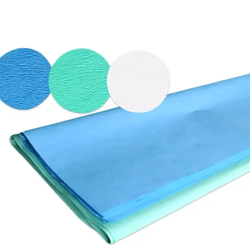
Crepe Paper for Sterile Safety Choosing the Right Medical Surgical Paper & Sterilization Wrapping Paper

Crepe paper isn’t just for crafts — in healthcare settings, high-quality Crepe Paper plays a vital role in protecting instruments and patients. From barrier performance to flexibility, modern creped materials deliver reliable breathability and tear resistance that make them ideal for surgical trays, instrument wraps, and packaging where sterility must be maintained without sacrificing handling convenience.


Hospitals and clinics increasingly prefer purpose-made Medical Surgical Paper because it balances microbial barrier properties with ease of use. Compared with plain non-woven sheets, specialty crepe constructions conform better around irregular instruments and give staff confident handling during sterilization cycles. When a wrap must pass autoclave or low-temperature sterilization while keeping contents sterile until point-of-use, choosing the right **Sterilization Wrapping Paper** directly affects instrument safety and workflow efficiency.


When evaluating suppliers look beyond paper weight and look for consistent crepe formation, controlled air permeability, and strong seam/edge performance — all features that reduce pinholes and maintain sterilization integrity. Practical considerations such as customizable roll widths, printed indicators, and compatibility with different sterilization methods help you match material to your facility’s protocols. Reliable sampling and clear technical data sheets speed adoption and reduce trial-and-error in procurement.


With more than 20 years as a factory serving global markets, Telijie combines proven product quality with service excellence: rapid sampling, flexible minimum orders, multilingual customer support, and tailored packaging and logistics to meet international delivery timelines. Beyond the material benefits of our Crepe Paper, Medical Surgical Paper, and **Sterilization Wrapping Paper**, Telijie offers hands-on technical guidance, responsive after-sales support, and the supply-chain consistency healthcare providers need—making Telijie the partner hospitals trust.

Top

Disposable Apron Buying Guide Why Dental & Paper Aprons Matter for Everyday Hygiene

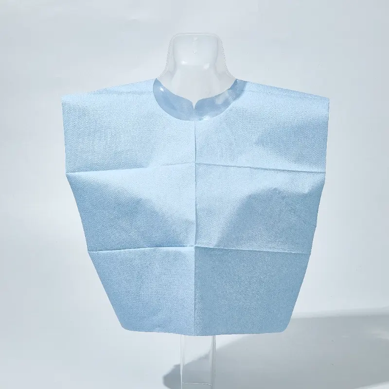
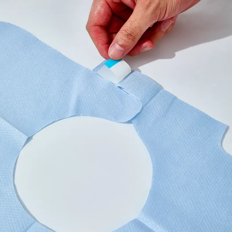
In environments where cleanliness and quick turnover matter, a reliable Disposable Apron is no longer optional — it’s essential. Whether you’re running a busy dental clinic, a food-prep station, or a light-industrial line, the right apron protects staff and strengthens your hygiene protocols without adding laundering costs or storage hassles.


Disposable aprons come in many forms to fit different needs. A Dental Apron is designed for splash protection, patient comfort and easy disposal after each procedure; lightweight but effective barriers reduce cross-contamination risk. For lightweight, single-use protection in salons, catering or quick medical checks, a Paper Apron offers an economical, biodegradable option that still delivers a clean-looking, professional presentation. Choosing the correct material and size ensures staff comfort while maintaining practical protection and reducing waste management burdens.


Beyond material choice, practical benefits include rapid restocking, consistent single-use hygiene, and simplified compliance for busy teams. Many businesses prefer disposable options because they eliminate laundry cycles, lower infection risk, and free up staff time for care rather than cleaning. When comparing alternatives, consider thickness, tie/neck style, and compatibility with other PPE so the Disposable Apron integrates seamlessly into your workflow.


For customers who require reliable supply and thoughtful service, Telijie stands apart. With over 20 years as a manufacturing partner serving global markets, Telijie combines factory-direct pricing with attentive OEM support, fast sample turnaround, and strict quality control throughout production. We help customers choose the right Disposable Apron, Dental Apron, or Paper Apron for their application, offer flexible customization (sizes, printing, packaging), and manage logistics to ensure steady supply. If you value decades of production experience plus responsive after-sales service, Telijie is built to deliver.

Top

Soft, Hygienic, Reliable Why Your Clinic Needs an Examination Couch Paper Roll

Clean, comfortable patient care begins with the small details — like the sheet that lies between patient and couch. For clinics, physiotherapy centers, and beauty salons that value hygiene and professionalism, the right paper roll makes a measurable difference to patient confidence and infection control. A high-quality Examination Couch Paper Roll protects surfaces, reduces laundering costs, and makes turnover between patients faster and neater.


Modern facilities need more than just a roll of paper; they need a dependable solution that performs under real-world pressure. Our Disposable Paper Bedsheet Roll options are designed for tear resistance, consistent unwind, and a smooth finish that won’t irritate skin. Whether you run a busy family practice or a specialist clinic, choosing the correct weight and width means fewer interruptions and a better experience for both staff and patients.


For medical settings where compliance and convenience matter most, Medical Couch Rolls combine sanitary performance with cost efficiency. Look for rolls manufactured under strict quality controls, available in perforated and non-perforated formats, and compatible with standard couch dispensers. These rolls simplify inventory management while supporting infection control protocols — a small change that yields big operational benefits.


As a factory with over 20 years’ experience serving the global market, Telijie knows the standards clinics expect. Beyond durable products, Telijie offers flexible OEM options, low minimum order quantities for trials, rapid production lead times, full quality inspection, and dependable international logistics. Our dedicated customer support helps with product selection, custom printing, and after-sales service — all designed to make switching to better Examination Couch Paper Roll and Medical Couch Rolls seamless for your practice. Choose Telijie for trusted products and service that keep your clinic running smoothly.

Top

Test Sooner, Act Faster – Enhance Sepsis Care with Poclight Diagnostics

1. The Global Burden of Sepsis


Sepsis is a life-threatening condition caused by the body’s extreme response to infection.

From data published in 2020, globally, over 49 million people are affected each year, with approximately 11 million deaths, representing 20% of all global deaths (Global, regional, and national sepsis incidence and mortality, 1990-2017: analysis for the Global Burden of Disease Study. Lancet. 2020 Jan 18).


sepsis worldwide


Sepsis affects people globally, but incidence and mortality are highest in lower-middle-income countries (LMICs). In high-income countries, the average hospital cost per sepsis patient exceeds US$32,000.


Understanding the Risk

Anyone can develop an infection that leads to sepsis, but certain factors increase susceptibility. Older adults, infants (40% of all cases), pregnant women, and patients with pre-existing conditions—such as chronic kidney disease, liver disease, cancer, or heart failure—face higher risk.


sepsis in children

early diagnosis of sepsis


Socioeconomic factors, access to healthcare, and living conditions also play a role, particularly in low- and middle-income countries where infections may be more prevalent and timely medical care is harder to access.

● High-risk populations: older adults, infants (half of all cases), pregnant women

● Pre-existing conditions: chronic kidney disease, liver disease, cancer, heart failure

● Socioeconomic & environmental factors: limited healthcare access, overcrowded living, poor sanitation


2. The Role of Biomarkers in Sepsis Diagnosis


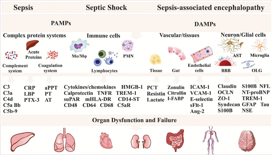
Inflammatory biomarkers are measurable indicators of the body’s immune response to infection. In sepsis, key inflammatory biomarkers may include the following:

sepsis biomarkers

Sepsis, septic shock, and sepsis-associated encephalopathy biomarkers. (Barichello, T., Generoso, J.S., Singer, M. et al. Biomarkers for sepsis: more than just fever and leukocytosis—a narrative review. Crit Care 26, 14 (2022))


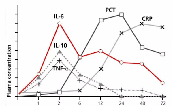
kinetics of key inflammation markers

Biomarker Kinetics After Bacterial Infection


Clinical benefits of monitoring these biomarkers:

● Early detection of sepsis and assessment of severity

● Informed decisions on initiating, adjusting, or discontinuing antimicrobial therapy

● Support antimicrobial stewardship (AMS): Reduced unnecessary antibiotic exposure without increasing mortality

● Real-time monitoring of treatment effectiveness alongside pathogen identification and antibiotic susceptibility testing


3. Poclight’s Solution: Speed, Precision, and Accessibility


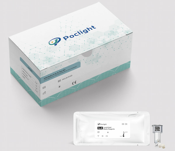
Poclight’s C5000 dry chemiluminescence immunoassay system offers a reliable approach to testing a panel of inflammatory and sepsis-related biomarkers, including PCT, CRP, IL-6, and HBP.

Category

Test

Sample types

Assay duration

Sample volume

Cut-Off value of Sepsis

Measuring range

INFLAMMATION

CRP test kit

Serum/Plasma

/Whole Blood

3min

5 μL

>100 mg/L

>10 mg/L neonatal

(low specificity, cannot diagnose sepsis alone)

0.5~320 mg/L

IL-6 test kit

Serum/Plasma

15min

100 μL

>250 pg/mL possible sepsis

1.5~5000 pg/mL

PCT test kit

Serum/Plasma

5min

200 μL

>0.5 ng/mL

high risk of sepsis

0.02~100 ng/mL

HBP test kit

Plasma

5min

50 μL

>28.1 ng/mL

5.9~300 ng/mL

Note: The reference values provided for each biomarker are for guidance only. Final clinical diagnosis should not rely on a single parameter, but instead be based on a comprehensive assessment that combines multiple biomarkers, diagnostic tools, and clinical evaluation.


1)  High Sensitivity & Specificity

Fully homogeneous chemiluminescence technology enables precise detection of small-molecule hormones and inflammatory markers, minimizing false results.

2)  Single-Test Freeze-Dried Reagents

Each assay comes as a single-dose, freeze-dried reagent that is stable at room temperature for up to 18 months, simplifying storage and transport—ideal for hospitals, primary care clinics, and remote settings.

Poclight freeze-dried reagents


3)  Rapid Turnaround

Test results in as fast as 3 minutes, enabling fast clinical decisions in critical situations.

4)  Cost-Effective: Low per-test cost allows frequent testing, saving healthcare budgets.

5)  Versatile and User-Friendly

Compact, simplified instruments make the system suitable for diverse clinical environments, from GP, clinics, to labs.

C5000 analyzer


By enabling early and accurate sepsis detection, Poclight diagnostics support clinicians in providing timely treatment while avoiding unnecessary broad-spectrum antibiotic use.


Detect sepsis early, respond rapidly, and save lives - Care in Time with Poclight

Get in touch with our specialists to get started and discover how we can help.

Top