How Electrosurgical Units Make Surgery Safer and Recovery Faster

Electrosurgical units (ESUs), commonly known as electrosurgical equipment, are widely used in surgical procedures. By generating heat through high-frequency electric current, they enable precise tissue cutting and effective hemostasis. Due to their ease of use, rapid cutting, and reliable bleeding control, ESUs have become indispensable tools in operating rooms, particularly in general surgery, gynecology, and colorectal surgery [1]. With the advancement of medical technology, the performance of electrosurgical units continues to improve, significantly enhancing both safety and efficiency, and supporting minimally invasive surgery and faster postoperative recovery.


Compared with traditional scalpels, electrosurgical units can effectively reduce intraoperative blood loss, shorten surgery duration, and lower the risk of postoperative complications. Studies have shown that proper use of ESUs can significantly improve surgical outcomes. In one study involving 160 surgical patients, the observation group receiving care with standardized electrosurgical unit procedures had an average surgery time of 132.9 minutes, intraoperative blood loss of 43.7 mL, and nursing satisfaction of 95.1%, all of which were superior to the conventional care group (P<0.05) [2]. This indicates that scientific usage and comprehensive nursing support are key to maximizing the advantages of electrosurgical units.


Safe use of electrosurgical units relies heavily on standardized nursing coordination. Nursing staff should check equipment status, electrode placement, and power connections before surgery to ensure a safe operating environment; during surgery, they must closely monitor the patient’s vital signs and equipment performance, addressing any anomalies promptly [3]. For example, selecting the appropriate output power, preventing patient contact with metal surfaces, and using insulation protection can effectively prevent burns and other accidents, ensuring smooth surgical procedures.


The SEH80A electrosurgical unit from ShouLiang-med integrates multiple intelligent technologies to further enhance surgical safety and efficiency. The device offers 8 monopolar and 6 bipolar modes to meet various surgical needs; its intelligent tissue-sensing function can adjust output power in real time, reducing smoke and eschar formation; and the return electrode monitoring system ensures patient safety from multiple dimensions. Additionally, the SEH80A supports laparoscopic interfaces, is compatible with smoke evacuators, and integrates into modern operating rooms, making it suitable for a variety of complex surgical scenarios and reflecting the efficiency and flexibility of modern surgical equipment.


In summary, electrosurgical units play a crucial role in modern surgery due to their significant intraoperative advantages and integration with proper nursing care. The application of advanced devices like the SEH80A from Shouliang-med has significantly improved surgical precision and safety, effectively promoting minimally invasive surgery and faster patient recovery.


References:

[1] Yan Weidong, Fan Li, Chen Hua, et al. Analysis of Nursing Coordination for the Safe Use of Electrosurgical Units in the Operating Room [J]. Medical Frontiers, 2016, 6(31): 286-287.

[2] Xiao Yingmei. Application Effect of Nursing Coordination for Safe Use of Electrosurgical Units in Surgery [J]. Medical Equipment, 2021, 34(4): 130-132.

[3] Long Yanli, Du Lin. Nursing Coordination for Safe Use of Electrosurgical Units in the Operating Room [J]. Medical Equipment, 2019, 32(10): 146-147.

Top

Maintenance and Servicing of AGISEAL Electrosurgical Generator SL100M

With the evolution of modern surgical treatment concepts, the number of OR medical devices continues to increase. The function and performance of these devices directly impact surgeons' operational proficiency and condition, ultimately affecting surgical outcomes. Therefore, ensuring optimal device performance during use is crucial. Here, we outline key maintenance and servicing points for the ShouLiang-med advanced electrosurgical vessel sealer dividers system SL100M.


How to Properly Install and Maintain the SL100M?


Installation:


  • First, inspect the high-frequency surgical generator and accessories to ensure they are complete and undamaged. Do not use any damaged devices.


  • Secure the high-frequency surgical equipment to a stable surface, such as a table, platform, or cart.


  • When placing the unit, ensure at least 10 cm to 15 cm of clearance is maintained above, on the sides, and behind the unit to facilitate cooling. 


  • Connect one end of the power line to the power socket on the rear panel of the generator, and plug the other end into a grounded power outlet. Connect the foot switch connector to the foot switch socket on the rear panel of the generator. Connect the instrument plug to the instrument socket on the front panel of the generator. 


  • Press the power switch to turn on the generator. The green light will illuminate. Verify the following: the company logo appears on the screen, The power-on self-test process emits an audible sound.  


  • If the self-test is successful, the main interface will be displayed, and the generator is ready for normal use.


  • If the self-test fails, the display will show the corresponding fault code. Please refer to the "Common Faults and Troubleshooting" section of the manual or contact an engineer.


Cleaning and Disinfection:


  • Thoroughly scrub all surfaces of the generator, power cord, and foot switch with a mild detergent or disinfectant solution and a soft damp cloth.The device can withstand frequent cleaning without degradation of the housing or display quality.


  • Warning: Risk of electric shock. Before cleaning, turn off the power supply and unplug the generator to ensure the device is isolated from the main power source.


  • Note: Do not use abrasive cleaning or disinfectant solutions (such as formalin), solvents, or other materials that may scratch the panel or damage the device.


  • Note: The instrument is not completely sealed. Liquids must not seep into the unit. Do not place liquid containers on the generator, and avoid spraying cleaning solutions directly onto it, as this may damage it.


Servicing


  • It is recommended that a comprehensive inspection of the generator and all accessories be conducted annually by qualified full-time personnel. Adjust the system according to technical requirements to ensure it remains in optimal condition.


  • It is recommended to inspect the power cord before each use of the system, or at intervals recommended by your hospital. Check the power cord for exposed wires, cracked outer casing, frayed edges, or damaged plugs. Replace any damaged cords. 


  • Malfunction of internal components may cause the fuse to blow. If the wall outlet has power but the generator fails to complete the power-on self-test or stops working, replace the fuse of the device.


  • During maintenance, surface dirt such as dust and oil stains can be cleaned using the traditional method of wiping with a dry cloth.


  • Avoid severe vibrations, exposure to harmful gases, and liquid ingress.


  • Keep the surfaces clean. Refer to the cleaning methods described above.
Top

Lab-Quality Thrombosis Testing with Poclight D-Dimer POC CLIA

1.  Thrombosis: An Overlooked Threat
 

The International Society on Thrombosis and Haemostasis (ISTH) established World Thrombosis Day (WTD) on October 13 to raise awareness of the global health threat.

Every year, 1 in 4 people worldwide die from conditions related to thrombosis, the underlying cause of the top three cardiovascular killers: heart attack, stroke, and venous thromboembolism (VTE). - World Thrombosis Day website (2025)

Venous Thromboembolism (VTE)


According to WHO data (2001–2023), the global age-standardized mortality rate of pulmonary embolism fell from 3.49 to 2.42 per 100,000. However, rates are rising in some lower-middle-income regions, underscoring the need to strengthen diagnostic capacity. Rapid and accessible POC testing helps bridge this gap where laboratory resources are limited.


2.  D-Dimer Testing Essentials


D-dimer is a specific fibrinolysis marker and key laboratory indicator of thrombus formation and breakdown. It rises within 2 hours of clot formation, has a 7-8 hour half-life, and remains stable in blood samples for 24-48 hours. These characteristics ensure its crucial role in diagnosing and monitoring thrombotic diseases.

Its clinical applications may include the following:

● Early Detection of Thrombosis: D-dimer testing is widely used to help rule out venous thromboembolism (VTE) in patients with suspected thrombotic events, particularly when combined with clinical assessment. Rapid detection supports prompt management decisions.

● Guiding Further Assessment: Prompts additional imaging or diagnostic tests.

● Supporting Anticoagulation Decisions: Helps guide initiation, adjustment, or duration of anticoagulant therapy.


Test Combination


Item

Clinical Use / Recommendation

D-dimer Test Kit

Helps rule out suspected DVT or PE (when combined with clinical probability assessment)

D-dimer + NT-proBNP Test Kit

Assists in differentiating PE from heart failure in patients with acute dyspnea

D-dimer + NT-proBNP and/or Troponin I

Helps rule out PE and assess PE prognosis

Note: D-Dimer levels can rise due to infection, inflammation, surgery, or pregnancy. It should always be interpreted alongside clinical findings and imaging.


3.  Poclight D-Dimer Solutions


D-Dimer Test Kit

Highly cost-effective, Poclight Biotech’s point-of-care D-dimer Test Kit solutions deliver results within 5 minutes, supporting prompt assessment and care.


Our Stand-out features:


1)  Small Sample Volume & Rapid Results

Only 50 μL of serum or plasma needed, with 5-minute turnaround, ideal for fast clinical decisions.

Parameter

Sample type

Assay duration

Sample volume

Cut-Off value

Measuring range

D-dimer

Plasma

5min

50 μL

0.5 mg/L, Higher D-dimer levels suggest clotting activity

0.25~20 mg/L


2)  Freeze-dried reagents: no cold chain, 18 months shelf life, simplifying storage and transport.

3)  High accuracy and excellent precision (CV < 5%)


DDimer Performance


4)  Applicable with the C5000 POC CLIA Analyzer: 3-Step Operation, Maintenance-Free

C5000 Operation


Contact us to learn about local distributors and pricing options for Poclight D-dimer solutions.

Top

Disposable Paper Bedsheet Roll Hygienic Medical Couch Rolls for Clinics

A clean, comfortable exam experience starts with the little details — and a high-quality Disposable Paper Bedsheet Roll makes a big difference. Clinics, spas, and mobile healthcare teams rely on disposable sheets to maintain hygiene between patients while keeping setup and cleanup quick. Designed for single-use convenience, these rolls help reduce cross-contamination risks and improve workflow in busy exam rooms.


For practitioners who need reliable, professional coverings, options such as Medical Couch Rolls and the Examination Couch Cover Roll are engineered to fit standard couches and trolleys, tear cleanly at perforations, and provide a soft, skin-friendly barrier. Good rolls balance strength and softness, resist minor fluids, and are available in a range of widths and ply to suit consultations, procedures, and physiotherapy sessions — all while being lightweight to store and simple to replace between patients.


At our factory we combine practical design with production discipline. With over 20 years’ experience manufacturing disposable medical disposables and supplying the global market, we offer OEM/ODM flexibility, consistent quality control, and competitive lead times. Customers benefit from tailored sizing, custom printing, and small-batch sampling so you can trial products before committing to larger runs. Our teams handle logistics and documentation for international shipping, making it straightforward to stock exam rooms anywhere in the world.


Choose Telijie when you want more than a product: you get a partner. Telijie’s strengths include stable, in-house manufacturing capacity, rigorous inspection at every stage, and dedicated account managers who respond quickly to orders, samples, and after-sales requests. We support healthcare buyers with clear packaging, compliant production practices, and flexible service options to match your inventory and budget needs — helping clinics and distributors maintain high standards without hassle.

Top

Revolutionizing Platelet Function Testing with Homogeneous CLIA Technology

1.What is Platelet?

Platelets, fragments of cytoplasm derived from bone marrow megakaryocytes, play a central role in hemostasis and thrombosis. When blood vessels are injured, platelets adhere, activate, and aggregate to stop bleeding. Abnormal platelet function can lead to bleeding disorders or thrombotic events. With the widespread use of antiplatelet drugs, such as aspirin and clopidogrel, precise assessment of platelet activation function is increasingly important. Poclight’s next-generation Homogeneous CLIA (Chemiluminescence Immunoassay) technology brings a new level of accuracy and speed to platelet function testing.

.What Are Platelets


2.Platelet Function and Clinical Significance

Platelet-mediated hemostasis involves three key steps:

a. Adhesion

When the endothelium is disrupted, platelets adhere to the vessel wall via von Willebrand Factor (VWF) and collagen. This process is mediated by glycoprotein receptors CD42a/b/d (GPIb-IX-V), a critical step in platelet adhesion.

b. Activation

Platelets are activated through G-protein-coupled receptors binding to ligands such as thrombin, ADP, ATP, and prostanoids. Activated platelets change shape to a dendritic or octopus-like form.

c. Aggregation

Activated platelets are interconnected by fibrinogen and receptors CD41/61 (GPIIb/IIIa). Platelet granules release their contents into the extracellular fluid, among which CD62P (P-selectin) promotes platelet aggregation through platelet–platelet and platelet–fibrin interactions.


Platelet function

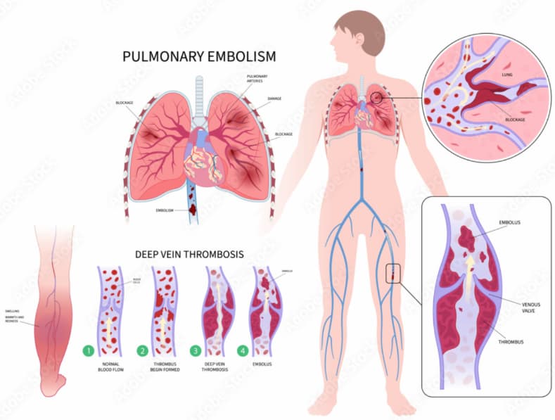
Hemostasis and thrombosis: The Key Role of Platelets 

Physiological function: The biological behavior and function of platelets are extremely complex. This includes participating in hemostasis and coagulation, maintaining vascular function, regulating inflammation, the body's immune defense, wound healing, tumor growth and metastasis, etc. Therefore, in clinical activities, the monitoring of platelet functionality is receiving increasing attention.

The average lifespan of platelets is 7 to 9 days. Senescent and dead platelets are eventually phagocytosed and processed in the spleen through the bloodstream.


Clinical Significance

  1. Thrombosis Risk Assessment:  Patients with Hypertension, diabetes,coronary, artery disease,and other cardiovascular condition, as well as those on antiplatelet or anticoagulant therapy.
  2. Pre/Postoperative Evaluation: Predicting thrombus formation and guiding prophylactic measures.
  3. Platelet Dysfunction Disorders: For example, Glanzmann’s thrombasthenia (CD41/61 deficiency) and Bernard-Soulier syndrome (CD42a/b/d deficiency).
  4.  Antiplatelet Therapy Monitoring: Supporting decisions for drugs like clopidogrel, ticagrelor, and prasugrel.
  5. Various detection functions:


Defect of platelet momoprotein receptor

CD61(GP IIIa)

CD(GP IIb)

CD42a(GP I X)

CD42b(GP I b)


Platelet Activation

CD62P(P

selectin)

CD61(GP IIIa)

CD61

Antibody(PAC-1)

CD45(White blood cell platelet aggregation)

Platelet Function

AA-CD62P

ADP-CD62P


3. Advantages of Poclight Platelet Activation Function Test

Feature

Description

Accurate & Fast

Quantitative results in just 5 minutes, enabling rapid clinical decisions.

Portable & Minimalist

One-step sample addition, maintenance-free, suitable for point-of-care testing.

True & Reliable

Technical details to be provided by the team to support this claim.

Quality Assurance

Supports quality control products and continuous QC checks, ensuring reliable results.

reagent testing

Technical Highlights:

Employs Homogeneous CLIA technology to detect multiple platelet surface markers (CD41, CD42, CD61, CD62P) for comprehensive assessment.

Evaluates the full spectrum of platelet function—adhesion, activation, and aggregation—delivering more comprehensive and physiologically-relevant results.


4. Compatible Devices

The Poclight Platelet Activation Function Test is fully compatible with Poclight latest C5000 Analyzer, offering seamless integration into clinical and point-of-care settings. Key compatibility features include:

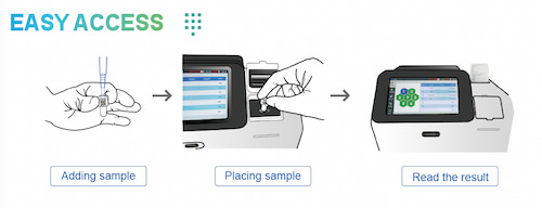
  1. Plug-and-Play Operation: Simple one-step sample addition for immediate testing.
  2. Portable Analyzer Integration: Works with compact, maintenance-free devices suitable for outpatient clinics or bedside monitoring.
  3. Future Expansion: Designed to support upcoming analyzer models based on the Homogeneous CLIA platform.
  4. Note: For detailed technical specifications and compatibility with your laboratory instruments, please contact our technical support team at overseas@poclight.com.

 Chemiluminescence immunoassay solution


5. Clinical Applications

  1. Outpatient Thrombosis Risk Screening: Quickly assess high-risk patients and guide antiplatelet therapy.
  2. Pre/Postoperative Monitoring: Track platelet activity changes to optimize surgical management.
  3. Antiplatelet Drug Efficacy Evaluation: Support individualized medication decisions and reduce bleeding or thrombotic events.
  4. Diagnosis of Platelet Dysfunction Disorders: Provide molecular-level insights for rare platelet diseases.


6.Influencing factors

  1. Genetic factors: Genes determine each person's basic platelet activity and their response to drugs.
  2. Drugs: Antiplatelet drugs, NSAIDs (such as ibuprofen), certain antibiotics, antidepressants, etc., can all affect their function.
  3. Disease status: Chronic diseases such as diabetes, kidney disease, and autoimmune diseases are often accompanied by high platelet reactivity (easier to form blood clots).
  4. Lifestyle:

Diet: Omega-3 fatty acids, garlic, ginger, certain vitamins (such as VC, VE) may inhibit platelet function.

Smoking and drinking: Smoking activates platelets, while moderate drinking may inhibit them.

Physiological conditions: Stress, exercise, and aging can all increase platelet activity.


7.Future Outlook

Platelet activation function testing is evolving towards speed, portability, standardization, and intelligence. Poclight’s Homogeneous CLIA platform not only shortens testing time but also delivers reliable, high-quality data, supporting precise assessment of bleeding and thrombotic risk. With ongoing technological innovation, this approach will play an increasingly vital role in precision medicine and individualized patient care.


8.Chemiluminescence resonance energy transfer(CRET)

Detection principle:

CRET cell technique utilizes the molecules on the platelet membrane to specifically fix and bind to the corresponding antibody 1-labeled DNA1, antibody 2-labeled DNA2, and AE-labeled DNA3 to form complexes. By detecting the light intensity via CRET, the expression levels of various antigens and receptor molecules in the sample can be detected.

Project Reference Interval Project

Reference Interval

CD62P

1.04-4.52%

CD42a

82.65-100%

CD61抗体

0-10%

CD42b

81.86-100%

CD61

60-92.13%

CD45

12.95%-46.04%

CD41

90.7-99.0%


Detection method:

  1. Nucleic acid technology is integrated into immunodiagnosis, leveraging the proximity efffect of nucleic acids takes effect.
  2. The properties of graphene oxide are strategically utilized, eliminating the need for magnetic beads and their associated separation and washing steps, and simplifying the fluidic path.
  3. The introduction of enzymatic cycling amplification technology enhances detection sensitivity by 1 to2 orders of magnitude, improving overall performance while simultaneously simplifying the process.


Advantage:

CRET compensates for the drawbacks of flow cytometry and enables:

  1. Fast, reliable detection in just 5-10 minutes.
  2.  A simple and minimalist operational procedure.
  3. Low-cost testing with no hidden fees.
  4. Easy adoption in all levels of hospitals and departments.
  5. Accurate in vivo status: Not affected by platelet aggregation or in vitro activation issues. 


Contact Us

E-mail: overseas@poclight.com


Top

AGISEAL A Safe and Effective Choice for Weight-loss Surgery

Since the 1950s, the medical field has been dedicated to solving the "century-old problem" of obesity. In 1952, Swedish doctor Henrikson attempted to treat obesity through extensive small bowel resection. By 1966, a doctor in the United States, inspired by the significant weight loss of patients who had undergone total gastrectomy, designed the prototype of the gastric bypass procedure. Since then, weight loss surgery has continually evolved, from open surgery to minimally invasive techniques like sleeve gastrectomy and RYGB gastric bypass, significantly improving safety and effectiveness.


There are various types of weight loss and metabolic surgeries, but sleeve gastrectomy has become the most widely used procedure globally due to its simplicity and lower risk of complications. By reducing stomach volume and restricting digestion and absorption, it can achieve long-term stable weight control and also improve obesity-related chronic diseases such as hypertension, diabetes, and sleep apnea. This has led to the shift of weight loss surgery from being merely a weight loss procedure to becoming a standard treatment for metabolic diseases.


As the number of patients undergoing these procedures continues to grow, the patient profile has also become clearer. Patients undergoing weight loss surgery typically have high demands for surgical safety and low tolerance for complications. Many guidelines emphasize the proper use of energy-based devices, as the occurrence of complications like gastric leakage is closely related to them. Since the early use of electrosurgical instruments in surgeries, devices that convert electrical energy into heat to achieve hemostasis have gradually replaced traditional cold knife cutting and suturing tools. Current mainstream devices, such as high-frequency electrosurgical knives, ultrasonic knives, and large vessel sealing devices, offer excellent hemostasis, quick cutting, high safety, and effectiveness, making them essential tools in modern surgery.


AGISEAL is one of these devices. It is based on the bipolar mode of high-frequency electrosurgical knives, with intelligent updates and iterations to the main unit technology, and has undergone optimization to enlarge the instrument's jaw for sealing and cutting large-diameter blood vessels (below 7mm). This significantly reduces the complex operations and time required by traditional suturing, earning it the title of "advanced bipolar." AGISEAL has shown excellent application in weight loss and metabolic surgeries and has received widespread praise from clinical experts. For example, under precise energy control, it reaches the minimum heat level, ensuring good clinical outcomes while maximizing the avoidance of burns to the gastric wall and the gastric fundus HIS angle, thereby reducing complications such as gastric leakage and gastroesophageal reflux. It meets patients' treatment needs while safeguarding their prognosis and precisely matches the patient profile.


Moreover, it combines safe sealing, rapid cutting, non-invasive holding, and fine dissection in one device, which means there is no need to switch surgical instruments or perform suturing and ligation when handling the stomach's greater omentum and short gastric arteries. A single SL0844 can efficiently complete the task, significantly reducing the operational time, helping to save on surgery time, and improving the operational efficiency of medical institutions and clinical departments, ultimately enhancing overall benefits.


In conclusion, the AGISEAL series is the undisputed choice to ensure surgical safety and improve efficiency in weight loss surgery.

Top

Bipolar Coagulation Forceps Enabling Precise Manipulation and Accelerated Recovery in Hemorrhoidectomy

Hemorrhoids are one of the most common anorectal diseases in clinical practice, with primary symptoms including bleeding, pain, and even prolapse, significantly impacting patients' quality of life. For patients with severe hemorrhoids, surgical excision is an effective treatment. Traditional surgery often employs monopolar electrocautery, which is widely used but associated with issues such as significant thermal damage and slow postoperative recovery. In recent years, with advancements in minimally invasive surgical techniques, bipolar coagulation forceps have been increasingly adopted in hemorrhoidectomy, demonstrating notable advantages.


Most medical institutions in China still commonly use monopolar electrocautery for hemorrhoidectomy. This device generates high heat through high-frequency current to achieve tissue cutting and coagulation. Although relatively simple to operate, the current must pass through the patient’s body to form a circuit, resulting in a broader thermal spread that can cause additional damage to healthy tissue around the wound edges. This leads to noticeable postoperative pain, prolonged wound healing, and a relatively higher incidence of complications such as urinary retention and incisional edema [1].


In contrast, bipolar coagulation forceps confine the current to a minimal area between the two tips, eliminating the need for a circuit through the body. The energy is highly concentrated, with minimal thermal diffusion. While achieving precise hemostasis, it maximally preserves surrounding healthy tissue, making it particularly suitable for the perianal region with dense vascular distribution. Clinical studies have shown that using bipolar coagulation forceps for hemorrhoidectomy significantly reduces operative time, intraoperative blood loss, postoperative pain scores, and promotes wound healing. Additionally, the incidence of complications such as postoperative edema and urinary dysfunction is significantly lower compared to traditional electrocautery [2].


ShouLiang-med’s independently developed bipolar coagulation forceps integrate high-precision tip design and anti-adhesive coating technology, ensuring concentrated current and minimal thermal damage. They are particularly suitable for delicate procedures such as hemorrhoidectomy. The "coagulation-dissection simultaneously" function enhances anatomical efficiency and operational smoothness, providing a clear surgical field and significantly improving surgical safety and postoperative recovery quality.


The application of bipolar coagulation forceps in hemorrhoidectomy offers multiple advantages, including minimal damage, rapid recovery, and fewer complications, aligning with the principles of modern minimally invasive surgery and enhanced recovery after surgery. ShouLiang-med’s bipolar coagulation forceps, with their excellent performance, are a reliable choice in the field of anorectal surgery.


References:

[1] Zhang P. Application analysis of procedure for prolapse and hemorrhoids (PPH) in the treatment of hemorrhoids. Chinese Journal of Medical Guide, 2016, 18(5): 440-441.

[2] Song J F. Application of bipolar coagulation forceps in hemorrhoidectomy. Journal of Clinical Medical Literature, 2019, 6(83): 73-74.

[3] Chen J Z, Xiang D Z, Geng X L, et al. Efficacy comparison of modified procedure for prolapse and hemorrhoids versus traditional surgery in the treatment of moderate-to-severe hemorrhoids. Modern Journal of Integrated Traditional Chinese and Western Medicine, 2013, 22(16): 1759-1760.

[4] Dai H, Hu Q, Huang J T, et al. Evaluation of bipolar coagulation technology in anorectal surgery. Journal of Hunan University (Medical Sciences), 2017, 14(1): 127-129.

Top

LEEP Electrosurgical Excision Procedure An Efficient and Precise Treatment for Cervical Diseases

Chronic cervicitis, including cervical erosion, hypertrophy, polyps, etc., are common conditions in women of childbearing age. In some gynecological outpatient departments, the incidence can reach nearly 10%. Traditional treatment methods such as cryotherapy, laser, microwave, and medications have had some efficacy but often fail to achieve satisfactory results and have high recurrence rates. In recent years, the incidence of CIN (Cervical Intraepithelial Neoplasia) has been rising among women of reproductive age, which is a crucial stage in the progression to invasive cervical cancer. Early diagnosis and treatment can significantly reduce the incidence of cervical cancer. Some hospitals have now included cervical carcinoma in situ in the scope of LEEP electrosurgical excision.


LEEP electrosurgical excision has shown significant advantages in the treatment of cervical diseases. According to the clinical observation study "Clinical Observation of LEEP Electrosurgical Excision for Cervical Diseases in 276 Cases," published by Wu Shengjun and others in Chinese Journal of Maternal and Child Health Research, this technique uses a high-frequency electrosurgical knife to precisely excise the diseased tissue. The procedure causes minimal discomfort for patients, with only a small number experiencing mild lower abdominal pain or a burning sensation. The average blood loss is only 5-6 mL, and the surgery time is as short as 7.3 minutes, with no need for hospitalization, making it an outpatient procedure. Postoperative recovery is quick, with 96.4% of patients showing normal cervical morphology within 1-2 months, and the overall complication rate is low.


Furthermore, the LEEP procedure combines both diagnostic and therapeutic functions. The excised tissue is free of carbonization, making it suitable for pathological examination, which is especially beneficial for early intervention in CIN. Compared to traditional methods such as cryotherapy and laser, LEEP has a lower recurrence rate and can preserve the patient's fertility. It is effective for various lesions such as cervical erosion, polyps, condyloma acuminata, and submucosal fibroids, and is easy to operate with high safety. Although long-term outcomes, such as the risk of cervical canal stenosis, still require more data validation, existing research indicates that its clinical value for widespread use is significant.


ShouLiang-med is committed to providing safe and stable high-frequency surgical equipment and a variety of gynecological surgical instruments. Its self-developed High Frequency Generator feature multiple cutting and coagulation modes, which meet the cutting and coagulation needs of gynecological surgeries, further reducing surgery time and blood loss, thereby ensuring patient safety. The company also offers different specifications of LEEP electrodes to meet the needs of various surgical scenarios. The electrodes are made of high-quality tungsten alloy, which further reduces the risks associated with tissue adhesion and carbonization during the procedure. Shouliang Medical's instruments are recognized by clinicians for their excellent materials, good anti-adhesion properties, and precise cutting and coagulation effects. The company hopes to provide safer solutions for global patients and more efficient energy systems for medical institutions in the future.

Top

Understanding Optical Coherence Tomography Technology in One Article.

I. What is OCT? A Core Metaphor


Let’s understand OCT through a vivid analogy:

Imagine a fresh sandwich. A regular camera can only capture its top view (a two-dimensional photo). OCT, however, acts like a precise “optical scalpel.” It can slice the sandwich layer by layer without damage, capturing ultra-high-resolution images of each component (bread, vegetables, meat slices). These cross-sectional images are then reconstructed into a three-dimensional model.

In the eye, this “sandwich” is your retina—the light-sensitive tissue at the back of the eyeball. OCT generates cross-sectional images of the retina’s microscopic structure without any incisions or contact, achieving micrometer-level precision (1 micrometer = 0.001 millimeters)—far finer than a strand of hair!


II. How Does OCT Work?

OCT operates on a principle similar to ultrasound, but instead of sound waves, it uses near-infrared light.

  1. Emission: The device emits a safe, low-energy near-infrared light beam toward your eye.
  2. Reflection: This light passes through the eyeball and reflects back from different layers of the retina. Each tissue layer reflects light signals with distinct intensities and time delays.
  3. Analysis: The device’s core interferometer precisely measures the differences between these reflected signals and the reference light (“interference” phenomenon).
  4. Imaging: A computer analyzes and processes the countless captured light signal points, instantly constructing detailed 2D or 3D images.

    III. What Can OCT Detect? Which Eye Conditions Does It Diagnose?
    OCT is a powerful tool for examining the macula (the central part of the retina responsible for sharpest vision). With it, doctors can:
  5. Precisely measure thickness: Determine whether the retina has thickened (edema, hemorrhage) or thinned (atrophy).
  6. Identify minute lesions: Detect fluid accumulation, abnormal membranes, and microbleeds invisible to the naked eye.
  7. Monitor disease progression dynamically: Evaluate treatment efficacy and guide subsequent therapeutic decisions.

    It is primarily applied in the diagnosis and management of these major blinding eye diseases:
  8. Age-related macular degeneration (AMD)

    Wet AMD: OCT clearly visualizes abnormal subretinal vessels and fluid accumulation, serving as the gold standard for diagnosis and determining the need for intravitreal injections.

    Dry AMD: OCT monitors for macular thinning (geographic atrophy) and the deposition of drusen.
  9. Diabetic Retinopathy & Diabetic Macular Edema (DME)

    OCT is the most sensitive tool for detecting DME (intraretinal fluid accumulation), precisely guiding laser or anti-VEGF drug injection treatments while objectively evaluating their efficacy.
  10. Glaucoma

    OCT accurately measures the thickness of the retinal nerve fiber layer (RNFL). Glaucoma causes the RNFL to gradually thin like the growth rings of a tree. OCT can detect these changes early, before visual field defects occur, enabling early diagnosis, timely treatment, and lifelong follow-up for glaucoma.
  11. Vitreoretinal Interface Diseases

    Such as epiretinal membrane (a membrane growing over the retina’s surface, causing distorted vision) and macular hole (a “hole” forming in the center of the macula). OCT clearly displays the morphology and staging of lesions, serving as a key basis for determining surgical necessity.

    IV. What is the OCT examination experience like?
  12. Preparation: Typically requires no special preparation. Sometimes dilating eye drops are administered to enlarge the pupil for clearer imaging, resulting in light sensitivity and blurred near vision for several hours afterward.
  13. Procedure: Simply rest your chin and forehead on the instrument’s support to keep your head steady. Focus on the fixation point inside the device (possibly a flashing cursor). The device does not touch your eyes.
  14. Duration: Scanning each eye usually takes only a few minutes.
  15. Safety: OCT uses near-infrared light. It is radiation-free, non-invasive, and has no known side effects. It is very safe and can be repeated frequently.

    V. Cutting-Edge Technology: Enhanced OCT
    OCT technology continues to evolve:
  16. OCT Angiography (OCTA): This is a revolutionary advancement. Without requiring contrast agent injections, it non-invasively reveals the intricate retinal vascular network, greatly facilitating the diagnosis of vascular diseases.
  17. Wider, Deeper, Faster: Scanning coverage is broader, penetration is deeper, speed is faster, and image quality is higher.

    3. Artificial Intelligence (AI): AI algorithms are being applied to automatically analyze OCT images, assisting physicians in rapid disease screening and diagnosis. This holds immense potential for future application in large-scale health screenings.


    Also welcome to contact us, we are ZD Medical Inc.
    Tel : +86-187 9586 9515
    Email : sales@zd-med.com
    Whatsapp/Mobile : +86-187 9586 9515

Top

What is the relationship between the New Vein Finder and pediatric emergency care?

Core Connection: Addressing the Most Challenging Clinical Scenarios in Pediatric Emergency Care

Pediatric emergency settings represent one of the most valuable applications for venous imaging devices. This connection is primarily manifested in addressing the following core challenges:


1. Patient Specificity: Difficulties in Establishing Venous Access

Physiological Factors: Infants and children possess inherently small, fragile blood vessels and relatively thick subcutaneous fat layers, making vessels invisible to the naked eye and difficult to palpate.

Emotional Factors: Young patients often experience fear, crying, and non-compliance, causing vasoconstriction and further complicating puncture attempts.

Dehydration/Critical Illness: In emergency settings, dehydrated, shock-affected, or hypotensive children exhibit collapsed vessels, making traditional localization nearly impossible.


2. Environmental Specificity: High Pressure and Urgency

Time Pressure: In critical situations (e.g., status epilepticus, severe dehydration, sepsis), rapidly establishing an IV line for medication administration or fluid infusion is vital for saving lives. Each failed attempt delays treatment.

Compounding Stress: A child's distress and parental anxiety create immense psychological pressure on healthcare providers, potentially leading to technical errors and creating a vicious cycle: “The more unsuccessful attempts, the greater the tension; the greater the tension, the fewer successful attempts.”

new vein imaging device

How does the new vein imaging device transform pediatric emergency care?

This innovative technology uses near-infrared light to project real-time images of subcutaneous veins directly onto the skin surface, clearly revealing vessel location, trajectory, and branching patterns.

Its specific value in pediatric emergencies includes:


1. Significantly increases first-attempt success rates

This is the most critical metric. Studies show vein imaging devices can boost first-attempt success rates in pediatric patients by 20% to 50% or more.

Significance: Reduces patient distress, shortens access establishment time, and secures a valuable window for critical interventions.


2. Reduces overall puncture attempts

Average attempts decrease from 3-4 to 1-2, or even one successful attempt.

Significance:

- Minimizes trauma for children: Prevents them from becoming “needle pads,” reducing physical and psychological harm.

- Alleviates parental anxiety: Significantly boosts parents' trust in the medical team.

- Protects healthcare providers: Lowers work frustration and enhances job satisfaction.


3. Provides solutions for “difficult vein” patients

For obese children, those with darker skin tones, severe dehydration, or multiple chemotherapy treatments, vein imaging devices are nearly the only non-invasive, rapid tool for locating veins.


4. Optimizing Workflow and Enhancing Emergency Department Efficiency

Reduces the need to call senior nurses or anesthesiologists for central venous catheterization due to repeated puncture attempts.

Enables healthcare providers to treat more patients faster, alleviating emergency department congestion.


5. As a Communication and Reassurance Tool

For patients: Project images onto their hands or toys, turning the process into a “game” to distract and reduce fear.

For parents: Visually demonstrate vein locations, helping them understand the procedure's difficulty and necessity. This transforms them from bystanders into “allies,” strengthening communication and trust.


Also welcome to contact us, we are ZD Medical Inc.
Tel : +86-187 9586 9515
Email : sales@zd-med.com
Whatsapp/Mobile : +86-187 9586 9515

Top