From -80℃ Freezing to 60℃ Heat: Which Transport Bag to Choose for Different Biological Samples? (With Advance Selection Guide)
Last week, we received an urgent inquiry from a research institute client: "Nearly a third of our transported blood samples suffered from hemolysis when they reached the lab, costing us half a month of experimental data!" Upon investigation, the issue traced back to the choice of transport bag—they had used ordinary PE bags, which could neither resist temperature fluctuations during transit nor provide anti-hemolysis protection.
In biological sample transportation, "choosing the right bag" matters more than "choosing an expensive one." Different samples (blood, tissues, viral reagents) have entirely different temperature tolerances and biological characteristics, meaning their corresponding transport bags also vary significantly in material, layer count, and protective design. Today, we’ll share a practical selection guide using Advance’s core products (AI650 Bag, Lab Absorbent Pouches) to help you avoid sample loss.
I. Blood Samples: Anti-Hemolysis Is Critical—Ordinary PE Bags Are a No-Go!
1. The "Temperature Dead Zone" for Blood Samples
Blood samples (whole blood, serum, plasma) are extremely sensitive to temperature fluctuations:
- During refrigerated storage (2-8℃), temperatures exceeding 10℃ accelerate red blood cell rupture; temperatures below 0℃ cause freezing, and ice crystals pierce cell membranes, directly leading to hemolysis.
- Even for short-distance transport (4-6 hours), the temperature fluctuation range must not exceed ±2℃—otherwise, it will affect subsequent blood routine and biochemical test results.
A previous client used ordinary PE bags to transport whole blood; a brief cold chain failure in the truck (temperature rose to 12℃) resulted in hemolysis of all 20 samples, causing tens of thousands of yuan in experimental losses.
2. Mandatory Material and Packaging Requirements
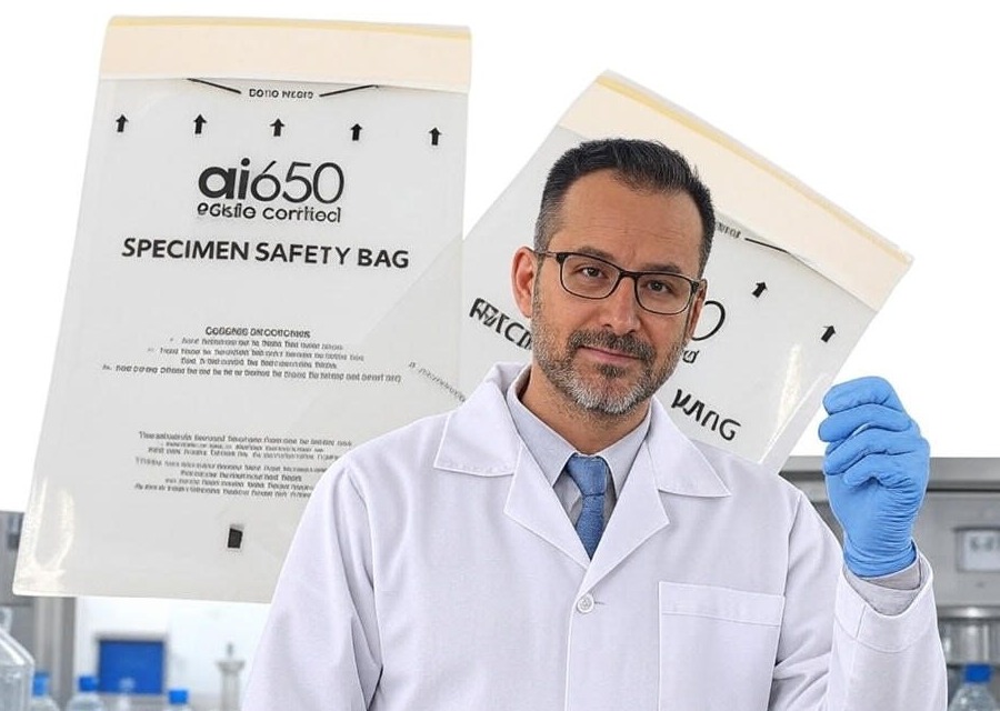
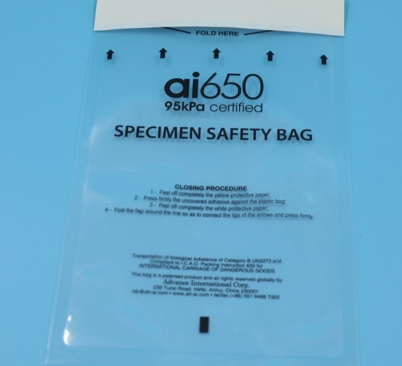
- Preferred Material: Five-Layer Co-Extruded Film (Core Material of AI650 Bag)
Ordinary PE bags have flaws: thin thickness (only 0.08mm), poor airtightness, easy oxygen permeation during temperature fluctuations, and potential leaching of trace additives that contaminate samples.
In contrast, Advance’s AI650 Bag uses a five-layer co-extruded film (LDPE/Tie/EVOH/Tie/ULDPE). The inner layer is medical-grade ULDPE with a leaching rate of less than 0.01%, avoiding reactions with proteins in blood. The middle EVOH layer has an oxygen transmission rate of only 0.3cc/(m²·24h)—90% lower than ordinary PE bags—locking in a stable internal environment and fundamentally reducing hemolysis risks.
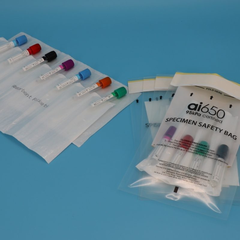
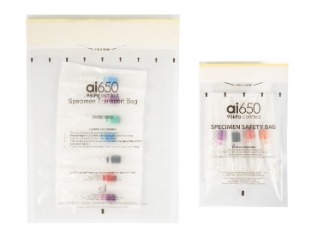
For even better results, pair it with Advance’s Lab Absorbent Pouches. This absorbent bag quickly soaks up blood that may accidentally leak due to seal issues (water absorption capacity up to 15x its own weight), preventing cross-contamination of samples while keeping the bag interior dry to further minimize temperature-induced impacts on blood.
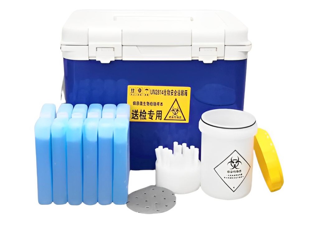
- Packaging Layers: At Least Double Layers; "AI650 Bag + Outer Cushion Bag"
Single-layer transport bags cannot withstand extrusion or vibration during transit. We recommend a double-layer structure: "inner AI650 Bag (for samples) + outer cushion bag (filled with bubble wrap)." In our tests, when whole blood-filled AI650 Bags were placed in cushion bags, the sample hemolysis rate was only 0.5% after a 1.2-meter drop test—far below the industry average of 3%.
3. Advance’s Test Data for Assurance
- 72-hour refrigerated transport at 2-8℃: Temperature fluctuation inside the AI650 Bag ≤ ±1℃, sample hemolysis rate < 1%.
- Accidental temperature rise to 30℃ (sustained for 2 hours): Thanks to the EVOH layer’s barrier, the internal temperature only rose to 12℃, not triggering the hemolysis threshold.
II. Tissue Samples: Low-Temperature Resistance + Puncture Protection—Even for -80℃ Freezing!
1. The "Preservation Challenge" for Tissue Samples
Tissue samples (e.g., tumor tissues, animal organs) typically require -80℃ freezing for preservation. During transport, it is necessary to maintain low temperatures and prevent sharp tissue edges from puncturing the bag. A client once used ordinary freezer bags to transport tumor tissues; the bag was pierced by tissue edges, and the sample deteriorated rapidly after exposure to air, forcing the experiment to be redone.
2. "Dual Protection" for Materials and Packaging
- Low-Temperature Resistant Material: AI650 Bag’s -196℃ Ultra-Low Temperature Tolerance
Ordinary freezer bags become brittle at -80℃ and crack easily when folded. However, the five-layer co-extruded film of Advance’s AI650 Bag undergoes low-temperature toughness treatment, remaining flexible even at -196℃ (liquid nitrogen temperature) with unchanged sealing performance after repeated folding.
For tissue samples that may leak bodily fluids, pairing with Lab Absorbent Pouches creates "dual protection": the absorbent bag soaks up leaked fluids, preventing them from freezing and puncturing the transport bag. Our clients report that this combination reduces tissue sample transport loss rates from 8% to below 1%.
- Packaging Layers: Three-Layer Protection for Extreme Transport Conditions
A three-layer structure is recommended: "inner AI650 Bag (for tissue samples) + middle Lab Absorbent Pouches (absorbent protection) + outer rigid carton (anti-extrusion)." Especially for frozen tissue transport, dry ice can be placed inside the carton. The AI650 Bag’s low-temperature resistance ensures samples are not damaged by freezing, while the outer carton prevents deformation caused by dry ice sublimation.
3. Advance’s Test Data for Assurance
After 72 hours of freezing at -80℃: The AI650 Bag maintains a 99.8% sealing rate. When the bag is opened, the tissue sample remains intact in shape with no freezing damage or deterioration.
Puncture resistance test: Pressing a sharp tissue edge (5mm diameter) against the AI650 Bag (50N pressure) for 10 minutes resulted in no damage or leakage.
III. Viral Reagents: Biosafety First, with Temperature Stability as a Priority
1. "Dual Risks" of Viral Reagents
Viral reagents (e.g., COVID-19, influenza virus samples) are not only temperature-sensitive (most require -20℃ freezing or 2-8℃ refrigeration) but also pose biosafety risks. If the transport bag is damaged, viral leakage could trigger public safety issues. Thus, compliance with UN Class 6.2 Dangerous Goods Transport Standards is mandatory.
2. "Compliant Design" for Materials and Packaging
Ordinary transport bags cannot meet the sealing requirements for dangerous goods transport. However, Advance’s AI650 Bag has passed the International Air Transport Association (IATA) Dangerous Goods Regulations (DGR) Edition 63 certification. Its double-seal design achieves 0 leakage, fully complying with biosafety transport standards for viral reagents.
Meanwhile, the absorbent layer of Lab Absorbent Pouches undergoes antibacterial treatment. Even if viral reagents accidentally leak, the antibacterial components in the absorbent bag inhibit viral activity and reduce spread risks—critical for cross-border transport (e.g., meeting EU IVDR regulatory biosafety requirements for exports).
- Packaging Layers: Designed by Hazard Level, At Least Three Layers
Packaging layers should be adjusted based on the risk level of viral reagents:
Low-risk viruses (e.g., ordinary influenza viruses): "AI650 Bag + Lab Absorbent Pouches + outer waterproof bag."
High-risk viruses (e.g., COVID-19): Add an outer rigid carton with biohazard labels to the above structure. The carton must pass a 1.2-meter multi-directional drop test (Advance’s custom cartons meet this requirement).
3. Advance’s Test Data for Assurance
96-hour refrigerated transport at 2-8℃: The activity retention rate of viral reagents inside the AI650 Bag reaches 95%—far higher than the industry average of 85%.
Biosafety test: Simulating a transport bag damage scenario, Lab Absorbent Pouches absorb all leaked reagents within 30 seconds, with an antibacterial rate of 99% and no viral spread risks.
|
Sample Type
|
Recommended Product Combination
|
Core Advantages
|
Transport Temperature Range
|
Notes
|
|
Blood Samples
|
AI650 Bag + Lab Absorbent Pouches
|
Anti-hemolysis, low leaching, minimal temperature fluctuation
|
2-8℃ (0-10℃ for short-term)
|
Avoid severe vibration; use with cushion bags
|
|
Tissue Samples
|
AI650 Bag + Lab Absorbent Pouches + Rigid Carton
|
Low-temperature resistance, puncture protection, stable sealing
|
-80℃ to 25℃
|
Use with dry ice for frozen transport; leave vent holes in carton
|
|
Viral Reagents
|
AI650 Bag + Lab Absorbent Pouches + Biosafety Carton
|
Compliant with Class 6.2 dangerous goods standards, antibacterial and leak-proof
|
-20℃ to 8℃
|
Attach biohazard labels to carton; include compliance documents with shipment
|
Is Your Sample Transport Still "Trial and Error"?
If your team is struggling with blood sample hemolysis, tissue sample freezing damage, or compliant transport of viral reagents, click here to visit www.aicbiologicalbag.com. Get a custom selection plan from Advance—we’ll recommend the most suitable AI650 Bag combination based on your sample type, transport distance, and temperature requirements. You can also request free samples for testing, ensuring "zero loss" in biological sample transport!